-
Sign In
-

-
 Sony Biotechnology
-

-
 Sony Biotechnology
PE anti-HMGB1
Antibodies Single
Sony
3E8
Flow Cytometry
Mouse IgG2b, κ
Human,Mouse,Rat
Recombinant human HMGB1 with GST tag. A universal T cell epitope from a Mycobacterium tuberculosis antigen was introduced into the C-terminus of HMGB1 to increase the immunogenicity.
3857020
$329.00
Description
High mobility group protein B1 (HMGB1) belongs to a family of highly conserved proteins that contain HMG box domains. Human HMGB1 is expressed as a 215 amino acid (aa) single chain polypeptide containing three domains: two N-terminal globular, 70 aa positively charged DNA binding domains (HMG boxes A and B), and a negatively charged 30 aa C-terminal region that contains only Asp and Glu. Human HMGB1 is 100% aa identical to canine HMGB1 and 99% aa identical to mouse, rat, bovine and porcine HMGB1.
HMGB1 is a widely expressed and highly abundant protein. It was originally discovered as a nuclear protein that could bend DNA. Such bending stabilizes nucleosome formation and regulates the expression of select genes upon recruitment by DNA binding proteins. It is now known that HMGB1 also plays a significant role in extracellular signaling associated with inflammation. HMGB1 is massively released into the extracellular environment during cell necrosis. It acts as an inflammatory mediator that promotes monocyte migration and cytokine secretion, and as a mediator of T cell-dendritic cell interaction. In addition, activated monocytes, macrophages, and dendritic cells also secrete HMGB1, forming a positive feedback loop that results in the release of additional cytokines and neutrophils. The cytokine activity of HMGB1 is restricted to the HMG B box, while the A box is associated with the helix-loop-helix domain of transcription factors. Although HMGB1 does not possess a classic signal sequence, it appears to be secreted as an acetylated form via secretory endolysosome exocytosis. Once secreted, HMGB1 transduces cellular signals through its high affinity receptor, RAGE and, possibly, TLR2 and TLR4.
Formulation
Phosphate-buffered solution, pH 7.2, containing 0.09% sodium azide and 0.2% (w/v) BSA (origin USA).Recommended Usage
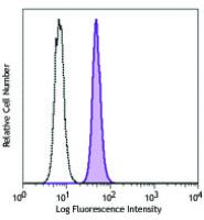
Each lot of this antibody is quality control tested by intracellular immunofluorescent staining using our nuclear factor staining protocol. For flow cytometric staining, the suggested use of this reagent is 5 microL per million cells or 5 microL per 100 microL of whole blood. It is recommended that the reagent be titrated for optimal performance for each application.
References
1. Zhou H, et al. 2009. PLos One 4:e6087. (Neut)


