


Brilliant Violet 750™ anti-mouse/human CD11b
Description
CD11b is a 170 kD glycoprotein also known as αM integrin, Mac-1 α subunit, Mol, CR3, and Ly-40. CD11b is a member of the integrin family, primarily expressed on granulocytes, monocytes/macrophages, dendritic cells, NK cells, and subsets of T and B cells. CD11b non-covalently associates with CD18 (β2 integrin) to form Mac-1. Mac-1 plays an important role in cell-cell interaction by binding its ligands ICAM-1 (CD54), ICAM-2 (CD102), ICAM-4 (CD242), iC3b, and fibrinogen.
Formulation
Phosphate-buffered solution, pH 7.2, containing 0.09% sodium azide and BSA (origin USA).Recommended Usage
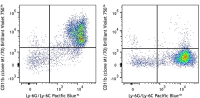
Each lot of this antibody is quality control tested by immunofluorescent staining with flow cytometric analysis. For flow cytometric staining, the suggested use of this reagent is ≤ 0.25 µg per million cells in 100 µl volume. It is recommended that the reagent be titrated for optimal performance for each application.
Brilliant Violet 750™ excites at 405 nm and emits at 750 nm. The bandpass filter 780/60 nm is recommended for detection, although filter optimization may be required depending on other fluorophores used. Be sure to verify that your cytometer configuration and software setup are appropriate for detecting this channel. Refer to your instrument manual or manufacturer for support. Brilliant Violet 750™ is a trademark of Sirigen Group Ltd.
This product is subject to proprietary rights of Sirigen Inc. and is made and sold under license from Sirigen Inc. The purchase of this product conveys to the buyer a non-transferable right to use the purchased product for research purposes only. This product may not be resold or incorporated in any manner into another product for resale. Any use for therapeutics or diagnostics is strictly prohibited. This product is covered by U.S. Patent(s), pending patent applications and foreign equivalents.
References
- Springer T, et al. 1978. Eur. J. Immunol. 8:539. (IP)
- Ault K and Springer T. 1981. J. Immunol. 126:359. (Deplete)
- Springer TA, et al. 1982. Immunol. Rev. 68:171. (Block)
- Ho MK and Springer TA. 1983. J. Biol. Chem. 258:2766. (IP)
- Flotte TJ, et al. 1983. Am. J. Pathol. 111:112. (IHC)
- Noel GJ, et al. 1990. J. Clin. Invest. 85:208. (IF)
- Allen LA and Aderem A. 1996. J. Exp. Med. 184:627 (IF)
- D'Amico A and Wu L. 2003. J. Exp. Med. 198:293. (Deplete)
- Brickson SJ, et al. 2003. Appl Physiol. 95:969. (Block)
- Clatworthy MR and Smith KG. 2004. J. Exp. Med. 199:717. (IF)
- Hata H, et al. 2004. J. Clin. Invest. 114:582. (IHC)
- Zhang Y, et al. 2002. J. Immunol. 168:3088. (IHC)
- Iwasaki A and Kelsall BL. 2001. J. Immunol. 166:4884 (IHC, FC)
- Tailleux L. 2003. J. Exp. Med. 197:121. (Block, FC)
- Olver S, et al. 2006. Cancer Research 66:571. (FC)
- Tan SL, et al. 2006. J. Immunol. 176:2872. (FC) PubMed
- Ponomarev ED, et al. 2006. J. Immunol. 176:1402. (FC)
- Dzhagalov I, et al. 2007. Blood 109:1620. (FC)
- Fazilleau N, et al. 2007. Nature Immunol. 8:753.
- Rasmussen JW, et al. 2006. Infect. Immun.74:6590. PubMed
- Napimoga MH, et al. 2008. J. Immunol. 180:609. PubMed
- Elqaraz-Carmon V, et al. 2008. J. Lipid. Res. 49:1894. PubMed
- Kim DD, et al. 2008. Blood 112:1109. PubMed
- Guo Y, et al. 2008. Blood 112:480. PubMed
- Norian LA, et al. 2009. Cancer Res. 69:3086. (FC) PubMed
- Baumgartner CK, et al. 2010. J. Immunol. 184:573. PubMed
- Charles N, et al. 2010. Nat. Med. 16:701. (FC) PubMed
- Whiteland J, et al. 1995. J. Histochem. Cytochem. 43:313. (IHC)
- Weber GF, et al. 2014. J Exp Med. 211:1243. PubMed
- Ashok A, et al. 2015. Toxicol Sci. 143:64. PubMed
- Price PJ, et al. 2015. J Immunol. 194:1164. PubMed
- Doni A, et al. 2015. J Exp Med. 212:905. PubMed
- Ferreira R, et al. 2016. J Infect Dis. 213: 669 - 673. PubMed
- Peterson VM, et al. 2017. Nat. Biotechnol. 35:936. (PG)