-
Sign In
-

-
 Sony Biotechnology
-

-
 Sony Biotechnology
Brilliant Violet 711™ anti-mouse/human CD44
Antibodies Single
Sony
IM7
Flow Cytometry
Rat IgG2b, κ
Human
Dexamethasone-induced myeloid leukemia M1 cells
50 µg
1115285
$295.00
Description
CD44 is a 80-95 kD glycoprotein also known as Hermes, Pgp1, H-CAM, or HUTCH. It is expressed on all leukocytes, endothelial cells, hepatocytes, and mesenchymal cells. As B and T cells become activated or progress to the memory stage, CD44 expression increases from low or mid levels to high levels. Thus, CD44 has been reported to be a valuable marker for memory cell subsets. High CD44 expression on Treg cells has been associated with potent suppressive function via high production of IL-10. CD44 is an adhesion molecule involved in leukocyte attachment to and rolling on endothelial cells, homing to peripheral lymphoid organs and to the sites of inflammation, and leukocyte aggregation.
Formulation
Phosphate-buffered solution, pH 7.2, containing 0.09% sodium azide and BSA (origin USA).Recommended Usage
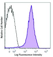
Each lot of this antibody is quality control tested by immunofluorescent staining with flow cytometric analysis. For flow cytometric staining, the suggested use of this reagent is ≤0.25 microg per million cells in 100 microL volume. It is recommended that the reagent be titrated for optimal performance for each application.
Brilliant Violet 711™ excites at 405 nm and emits at 711 nm. The bandpass filter 710/50 nm is recommended for detection, although filter optimization may be required depending on other fluorophores used. Be sure to verify that your cytometer configuration and software setup are appropriate for detecting this channel. Refer to your instrument manual or manufacturer for support. Brilliant Violet 711™ is a trademark of Sirigen Group Ltd.
References
1. Trowbridge IS, et al. 1982. Immunogenetics 15:299. (ICFC, IP, CMCD)
2. Katoh S, et al. 1994. J. Immunol. 153:3440. (ELISA)
3. Budd RC, et al. 1987. J. Immunol. 138:3120. (IP)
4. Camp RL, et al. 1993. J. Exp. Med. 178:497. (Block)
5. Weiss JM, et al. 1997. J. Cell Biol. 137:1137. (Block)
6. Frank NY, et al. 2005. Cancer Res. 65:4320. (IHC) PubMed
7. Cuff CA, et al. 2001. J. Clin. Invest. 108:1031. (IHC)
8. Lee JW, et al. 2006. Nature Immunol. 8:181.
9. Zhang N, et al. 2005. J. Immunol. 174:6967. PubMed
10. Huabiao C, et al. 2005. J. Immunol. 175:591. PubMed
11. Gui J, et al. 2007. Int. Immunol. 19:1201. PubMed
12. Wang XY, et al. 2008. Blood 111:2436. PubMed
13. Kenna TJ, et al. 2008. Blood 111:2091. PubMed
14. Yamazaki J, et al. 2009. Blood PubMed
15. Kmieciak M, et al. 2009. J. Transl. Med. 7:89. (FC) PubMed
16. Chen YW, et al. 2010. Mol. Cancer Ther. 9:2879. PubMed
17. Zheng Z, et al. 1995. J. Cell. Biol. 130:485.
18. Wiranowska M, et al. 2010. Int. J. Cancer 127:532.
19. Hirokawa Y, et al. 2014. Am J Physiol Gastrointerest Liver Physiol. 306:547. PubMed
20. Sandmaier BM, et al. 1998. Blood 91:3494.


