-
Sign In
-

-
 Sony Biotechnology
-

-
 Sony Biotechnology
Alexa Fluor® 700 anti-human CD3
Antibodies Single
Sony
OKT3
Flow Cytometry
Mouse IgG2a, κ
Human
2186700
$260.00
Description
CD3ε is a 20 kD chain of the CD3/T cell receptor (TCR) complex, which is composed of two CD3ε, one CD3γ, one CD3δ, one CD3ζ (CD247), and a T cell receptor (α/β or γ/δ) heterodimer. It is found on all mature T lymphocytes, NK T cells, and some thymocytes. CD3, also known as T3, is a member of the immunoglobulin superfamily that plays a role in antigen recognition, signal transduction, and T cell activation.
Formulation
Phosphate-buffered solution, pH 7.2, containing 0.09% sodium azide and 0.2% (w/v) BSA (origin USA).Recommended Usage
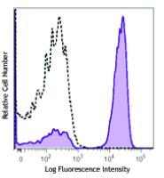
Each lot of this antibody is quality control tested by immunofluorescent staining with flow cytometric analysis. For flow cytometric staining, the suggested use of this reagent is 5 microL per million cells or 5 microL per 100 microL of whole blood. It is recommended that the reagent be titrated for optimal performance for each application.
* Alexa Fluor® 700 has a maximum emission of 719 nm when it is excited at 633 nm / 635 nm. Prior to using Alexa Fluor® 700 conjugate for flow cytometric analysis, please verify your flow cytometer's capability of exciting and detecting the fluorochrome.
References
1. Schlossman S, et al. Eds. 1995. Leucocyte Typing V. Oxford University Press. New York.
2. Knapp W. 1989. Leucocyte Typing IV. Oxford University Press New York.
3. Barclay N, et al. 1997. The Leucocyte Antigen Facts Book. Academic Press Inc. San Diego.
4. Li B, et al. 2005. Immunology 116:487.
5. Jeong HY, et al. 2008. J. Leuckocyte Biol. 83:755. PubMed
6. Alter G, et al. 2008. J. Virol. 82:9668. PubMed
7. Manevich-Mendelson E, et al. 2009. Blood 114:2344. PubMed
8. Pinto JP, et al. 2010. Immunology. 130:217. PubMed
9. Biggs MJ, et al. 2011. J. R. Soc. Interface. 8:1462. PubMed


