-
Sign In
-

-
 Sony Biotechnology
-

-
 Sony Biotechnology
FITC anti-human CD39
Antibodies Single
Sony
A1
Flow Cytometry
Mouse IgG1, κ
Human
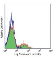
PHA activated human lymphocytes
2241030
$226.00
Description
Human CD39 is an integral membrane protein with two transmembrane domains. It exists as a homotetramer. Expression of CD39 is found on activated lymphocytes, a subset of T cells and B cells, and dendritic cells with weak staining on monocytes and granulocytes. CD39 and CD73 have been found on regulatory T cells, specifically the effector/memory like T cells. CD39 can hydrolyze both nucleoside triphosphates and diphosphates. CD39 is the dominant ecto nucleotidase of vascular and placental trophoblastic tissues and appears to modulate the functional expression of type 2 purinergic (P2) G protein coupled receptors (GPCRs). CD39 has intrinsic ecto-ATPase activity. Expression of CD39 is induced on T cells and increased on B cells as a late activation antigen.
Formulation
Phosphate-buffered solution, pH 7.2, containing 0.09% sodium azide and 0.2% (w/v) BSA (origin USA).Recommended Usage
Each lot of this antibody is quality control tested by immunofluorescent staining with flow cytometric analysis. Test size products are transitioning from 20 microL to 5 microL per test. Please check your vial or your CoA to find the suggested use of this reagent per million cells in 100 microL staining volume or per 100 microL of whole blood. It is recommended that the reagent be titrated for optimal performance for each application.
References
1. Aversa GG, et al. 1988. Transplant. P. 20:4952.
2. Aversa GG, et al. 1989. Transplant. P. 21:34950.
3. Borsellino G, et al. 2007. Blood. 110:1225. (Block)
4. Stockl J, et al. 2001. J. Immunol. 167:2724. (IF)
5. Sestak K, et al. 2007. Vet. Immunol. Immunopathol. 119:21.
6. Lyck L, et al. 2008. J. Histochem. Cytochem. 56:201. (IHC)


