-
Sign In
-

-
 Sony Biotechnology
-

-
 Sony Biotechnology
PerCP anti-mouse CD8a
Antibodies Single
Sony
53-6.7
Flow Cytometry
Rat IgG2a, κ
Mouse
Mouse thymus or spleen
1103655
$105.00
Description
CD8, also known as Lyt-2, Ly-2, or T8, consists of disulfide-linked α and β chains that form the α(CD8a)/β(CD8b) heterodimer and α/α homodimer. CD8a is a 34 kD protein that belongs to the immunoglobulin family. The CD8 α/β heterodimer is expressed on the surface of most thymocytes and a subset of mature TCR α/β T cells. CD8 expression on mature T cells is non-overlapping with CD4. The CD8 α/α homodimer is expressed on a subset of γ/δ TCR-bearing T cells, NK cells, intestinal intraepithelial lymphocytes, and lymphoid dendritic cells. CD8 is an antigen co-receptor on T cells that interacts with MHC class I on antigen-presenting cells or epithelial cells. CD8 promotes T cell activation through its association with the TCR complex and protein tyrosine kinase lck.
Formulation
Phosphate-buffered solution, pH 7.2, containing 0.09% sodium azide.Recommended Usage
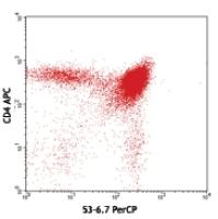
Each lot of this antibody is quality control tested by immunofluorescent staining with flow cytometric analysis. For flow cytometric staining, the suggested use of this reagent is ≤ 0.25 microg per 106 cells in 100 microL volume. It is recommended that the reagent be titrated for optimal performance for each application.
* PerCP has a maximum absorption of 482 nm and a maximum emission of 675 nm.
References
1. Ledbetter JA, et al. 1979. Immunol. Rev. 47:63. (IHC, IP)
2. Hathcock KS. 1991. Current Protocols in Immunology. 3.4.1. (Deplete)
3. Takahashi K, et al. 1992. P. Natl. Acad. Sci. USA 89:5557. (Block, IP)
4. Ledbetter JA, et al. 1981. J. Exp. Med. 153:1503. (Block)
5. Hata H, et al. 2004. J. Clin. Invest. 114:582. (IHC)
6. Fan WY, et al. 2001. Exp. Biol. Med. 226:1045. (IHC)
7. Shih FF, et al. 2006. J. Immunol. 176:3438. (FC)
8. Kamimura D, et al. 2006. J. Immunol. 177:306.
9. Bouwer HGA, et al. 2006. P. Natl. Acad. Sci. USA 103:5102. (FC, Deplete)
10. Kao C, et al. 2005. Int. Immunol. 17:1607. PubMed
11. Ko SY, et al. 2005. J. Immunol. 175:3309. (FC) PubMed
12. Rasmussen JW, et al. 2006. Infect. Immun. 74:6590. PubMed
13. Lee CH, et al. 2009. Clin. Cancer Res. PubMed
14. Geiben-Lynn R, et al. 2008. Blood 112:4585. (Deplete) PubMed
15. Kingeter LM, et al. 2008. J. Immunol. 181:6244. PubMed
16. Guo Y, et al. 2008. Blood 112:480. PubMed
17. Andrews DM, et al. 2008. J. Virol. 82:4931. PubMed
18. Britschqui MR, et al. 2008. J. Immunol. 181:7681. PubMed
19. Kenna TJ, et al. 2008. Blood 111:2091. PubMed
20. Jordan JM, et al. 2008. Infect. Immun. 76:3717. PubMed
21. Todd DJ, et al. 2009. J. Exp. Med. 206:2151. PubMed
22. Bankoti J, et al. 2010. Toxicol. Sci. 115:422. (FC) PubMed
23. Medyouf H, et al. 2010. Blood 115:1175. PubMed
24. Riedl P, et al. 2009. J. Immunol. 183:370. PubMed
25. Apte SH, et al. 2010. J. Immunol. 185:998. PubMed
26. Bankoti J, et al. 2010. Toxicol. Sci. 115:422. (FC) PubMed
27. del Rio ML, et al. 2011. Transpl. Int. 24:501. (FC) PubMed
28. Sun Z, et al. 2013. Biochem Biophys Res Commun. 435:472. PubMed
29. Brenndorfer ED, et al. 2014.J. Immunol. 192:1671. PubMed
30. Byrne KT, et al. 2014. J Immunol. 192:1433.


