


Brilliant Violet 750™ anti-mouse CD11c
Description
CD11c is a 150 kD glycoprotein also known as αX integrin, CR4, and p150. CD11c forms a αXβ2 heterodimer with β2 integrin (CD18). It is primarily expressed on dendritic cells, NK cells, a subset of intestinal intraepithelial lymphocytes (IEL), and some activated T cells. The αXβ2 integrin plays an important role in cell-cell contact by binding its ligands: iC3b, fibrinogen, and CD54.
Formulation
Phosphate-buffered solution, pH 7.2, containing 0.09% sodium azide and BSA (origin USA).Recommended Usage
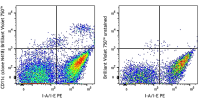
Each lot of this antibody is quality control tested by immunofluorescent staining with flow cytometric analysis. For flow cytometric staining, the suggested use of this reagent is ≤0.125 µg per million cells in 100 µl volume. It is recommended that the reagent be titrated for optimal performance for each application.
Brilliant Violet 750™ excites at 405 nm and emits at 750 nm. The bandpass filter 780/60 nm is recommended for detection, although filter optimization may be required depending on other fluorophores used. Be sure to verify that your cytometer configuration and software setup are appropriate for detecting this channel. Refer to your instrument manual or manufacturer for support. Brilliant Violet 750™ is a trademark of Sirigen Group Ltd.
This product is subject to proprietary rights of Sirigen Inc. and is made and sold under license from Sirigen Inc. The purchase of this product conveys to the buyer a non-transferable right to use the purchased product for research purposes only. This product may not be resold or incorporated in any manner into another product for resale. Any use for therapeutics or diagnostics is strictly prohibited. This product is covered by U.S. Patent(s), pending patent applications and foreign equivalents.
References
- Granucci F, et al. 1997. J. Immunol. 159:1794.
- Stokes RW, et al. 1998. J. Immunol. 160:5514.
- Metlay JP, et al. 1990. J. Exp. Med. 171:1753. (IHC, IP)
- Ma XT, et al. 2006. Cancer Research 66:1169.
- Chin RK, et al. 2006. J. Immunol. 177:290. (IF)
- Cervantes-Barragan L, et al. 2007. Blood 109:1131. (FC) PubMed
- Turnquist HR, et al. 2007. J. Immunol. 178:7018. (FC) PubMed
- Benson MJ, et al. 2007. J. Exp. Med. doi:10.1084/jem.20070719. (FC) PubMed
- You Y, et al. 2009. J. Immunol. 182:7343. (IF) PubMed
- Roland CL, et al. 2009. Mol. Cancer Res. 8:1761. (IHC, FC) PubMed
- Wikstrom M, et al.2006. J. Immunol. 177:913. PubMed
- Pericolini E, et al. 2008. J. Leukocyte Biol. 83:1286. PubMed
- Randall LM, et al. 2008. Infect. Immun.76:3312. PubMed
- Fahlen-Yrild L, et al. 2009. J. Immunol. 183:5032. PubMed
- Osterholzer JJ, et al. 2009. J. Immunol. 183:8044. PubMed
- Bankoti J, et al. 2010. Toxicol. Sci. 115:422. (FC) PubMed
- Eisenach PA, et al. 2010. J Cell Sci. 123:4182. PubMed
- Leppin K, et al. 2014. Invest. Ophthalmol. Vis. Sci. 55:3603. PubMed
- Sakai F, et al. 2014. PLoS One. 9:105370. PubMed
- Gibbins JD, et al. 2014. Blood. 124:2953. PubMed
- White CE, et al. 2015. J Immunol. 194:697. PubMed
- Lu X, et al. 2015. J Immunol. 194:2011. PubMed