


Brilliant Violet 605™ anti-human CD28
Description
CD28 is a 44 kD disulfide-linked homodimeric type I glycoprotein. It is a member of the immunoglobulin superfamily and is also known as T44 or Tp44. CD28 is expressed on most T lineage cells, NK cell subsets, and plasma cells. CD28 binds both CD80 and CD86 using a highly conserved motif MYPPY in the CDR3-like loop. CD28 is considered a major co-stimulatory molecule, inducing T lymphocyte activation and IL-2 synthesis, and preventing cell death. In vitro studies indicate that ligation of CD28 on T cells by CD80 and CD86 on antigen presenting cells provides a costimulatory signal required for T cell activation and proliferation.
Recommended Usage
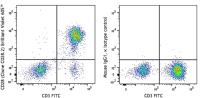
Each lot of this antibody is quality control tested by immunofluorescent staining with flow cytometric analysis. For flow cytometric staining, the suggested use of this reagent is 5 µl per million cells in 100 µl staining volume or 5 µl per 100 µl of whole blood.
Brilliant Violet 605™ excites at 405 nm and emits at 603 nm. The bandpass filter 610/20 nm is recommended for detection, although filter optimization may be required depending on other fluorophores used. Be sure to verify that your cytometer configuration and software setup are appropriate for detecting this channel. Refer to your instrument manual or manufacturer for support. Brilliant Violet 605™ is a trademark of Sirigen Group Ltd.
This product is subject to proprietary rights of Sirigen Inc. and is made and sold under license from Sirigen Inc. The purchase of this product conveys to the buyer a non-transferable right to use the purchased product for research purposes only. This product may not be resold or incorporated in any manner into another product for resale. Any use for therapeutics or diagnostics is strictly prohibited. This product is covered by U.S. Patent(s), pending patent applications and foreign equivalents.
References
- Schlossman S, et al. Eds. 1995. Leucocyte Typing V. Oxford University Press. New York.
- Nunes J, et al. 1993. Biochem. J. 293:835.
- Calea-Lauri J, et al. 1999. J. Immunol. 163:62.
- Tazi A, et al. 1999. J. Immunol. 163:3511. (IHC)
- Marti F, et al. 2001. J. Immunol. 166:197. (Costim)
- Jeong SH, et al. 2004. J. Virol. 78:6995. (Costim)
- Rivollier A, et al. 2004. Blood 104:4029. (Costim)
- Scharschmidt E, et al. 2004. Mol. Cell Biol. 24:3860. (Costim)
- Sheng W, et al. 2007.Elsevier 580:6819. PubMed
- Mitsuhashi M. 2007. Clin Chem.53:148. PubMed
- Ye Z, et al. 2008. Infect. Immun. 76:2541. PubMed
- Magatti M, et al. 2008. Stem Cells 26:182. (FA) PubMed
- Yoshino N, et al. 2008. Exp. Anim. (Tokyo) 49:97. (FC)
- Berg M, et al. 2008. J Leukoc Biol. 83:853. (IP) PubMed
- Rout N, et al. 2010. PLoS One 5:e9787. (FC)
- Leonard JA, et al. 2011. J. Virol. 85:6867. PubMed
- Nomura T, et al. 2012. J. Virol. 86:6481. PubMed