


Brilliant Violet 785™ anti-human CD2
Description
CD2 is a 50 kD type I transmembrane glycoprotein also known as LFA-2, T11, and sheep red blood cell receptor (SRBC-R). This immunoglobulin superfamily member is expressed on thymocytes, T lymphocytes, NK cells, and thymic B cell subsets. The major ligand for CD2 is CD58 (also known as LFA-3). CD2 has also been reported to bind CD48, CD59, and CD15. CD2 plays a critical role in alternative T cell activation, T cell signaling, and cell-cell adhesion.
Formulation
Phosphate-buffered solution, pH 7.2, containing 0.09% sodium azide and BSA (origin USA).Recommended Usage
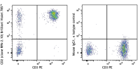
Each lot of this antibody is quality control tested by immunofluorescent staining with flow cytometric analysis. For flow cytometric staining, the suggested use of this reagent is 5 µl per million cells in 100 µl staining volume or 5 µl per 100 µl of whole blood.
Brilliant Violet 785™ excites at 405 nm and emits at 785 nm. The bandpass filter 780/60 nm is recommended for detection, although filter optimization may be required depending on other fluorophores used. Be sure to verify that your cytometer configuration and software setup are appropriate for detecting this channel. Refer to your instrument manual or manufacturer for support. Brilliant Violet 785™ is a trademark of Sirigen Group Ltd.
This product is subject to proprietary rights of Sirigen Inc. and is made and sold under license from Sirigen Inc. The purchase of this product conveys to the buyer a non-transferable right to use the purchased product for research purposes only. This product may not be resold or incorporated in any manner into another product for resale. Any use for therapeutics or diagnostics is strictly prohibited. This product is covered by U.S. Patent(s), pending patent applications and foreign equivalents.
References
- Knapp W, et al. Eds. 1989. Leucocyte Typing IV. Oxford University Press. New York.
- Aversa G, et al. 1987. Transplant. Proc. 19:277. (Block)
- Zaretsky AG, et al. 2009. J. Exp Med. 206:991. (IHC) PubMed
- Perona-Wright G, et al. 2010. Nat. Immunol. 11:520. (FC) PubMed
- Thummler K, et al. 2010. J. Leukoc. Biol. 88:1041.
- Kap Y, et al. 2009. J. Histochem. Cytochem. 57:1159. (IHC)
- Yoshino N, et al. 2000. Exp. Anim. (Tokyo) 49:97. (FC)