-
Sign In
-

-
 Sony Biotechnology
-

-
 Sony Biotechnology
Alexa Fluor® 647 anti-mouse CD4
Antibodies Single
Sony
GK1.5
Immunofluorescence
Rat IgG2b, κ
Mouse
Mouse CTL clone V4
1102130
$98.00
Description
CD4 is a 55 kD protein also known as L3T4 or T4. It is a member of the Ig superfamily, primarily expressed on most thymocytes, a subset of T cells, and weakly on macrophages and dendritic cells. It acts as a coreceptor with the TCR during T cell activation and thymic differentiation by binding MHC class II and associating with the protein tyrosin kinase, lck.
Formulation
Phosphate-buffered solution, pH 7.2, containing 0.09% sodium azide.Recommended Usage
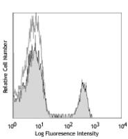
Each lot of this antibody is quality control tested by immunofluorescent staining with flow cytometric analysis. For flow cytometric staining, the suggested use of this reagent is ≤ 0.25 microg per million cells in 100 microL volume. For immunohistochemisty, a concentration range of 2.5-5 μg/ml is suggested. For immunofluorescence microscopy, a concentration range of 1.25-10 μg/ml is recommended. It is recommended that the reagent be titrated for optimal performance for each application.
* Alexa Fluor® 647 has a maximum emission of 668 nm when it is excited at 633nm / 635nm.
References
1. Dialynas DP, et al. 1983. J. Immunol. 131:2445. (Block, IP)
2. Dialynas DP, et al. 1983. Immunol. Rev. 74:29. (IP, Deplete)
3. Wu L, et al. 1991. J. Exp. Med. 174:1617. (Costim)
4. Godfrey DI, et al. 1994. J. Immunol. 152:4783. (Block)
5. Gavett SH, et al. 1994. Am. J. Respir. Cell. Mol. Biol. 10:587. (Deplete)
6. Schuyler M, et al. 1994. Am. J. Respir. Crit. Care Med. 149:1286. (Deplete)
7. Ghobrial RR, et al. 1989. Clin. Immunol. Immunopathol. 52:486. (Deplete)
8. Israelski DM, et al. 1989. J. Immunol. 142:954. (Deplete)
9. Zheng B, et al. 1996. J. Exp. Med. 184:1083. (IHC)
10. Frei K, et al. 1997. J. Exp. Med. 185:2177. (IHC)
11. Felix NJ, et al. 2007. Nat. Immunol. 8:388. (Block)
12. Li J, et al. 2012. Arthritis Rheum. 64:1098. PubMed
13. Ishida W, et al. 2014. Cell Immunol. 153:136. PubMed


