


-
Cancer immunotherapies: How single-cell sequencing can aid informed decision-making

An insider’s view on leveraging the latest single cell technologies including cell sorting and single-cell sequencing to understand what drives response or resistance in checkpoint therapies.

Moshe Sade Feldman (PhD) is an Instructor in Medicine and the Director of the Translational Cancer Immunology lab at Massachusetts General Hospital Cancer Center. Immune checkpoint blockade (ICB) therapies have revolutionized oncology treatment and become the standard of care in treating malignancies such as solid tumors1,2. Despite proving more efficient than conventional chemotherapies or targeted therapies, due to their relatively high rate of response (up to 45% in melanoma patients), ICB presents challenges such as treatment resistance and eventual relapse. As cancer treatments evolve, some of the biggest unknowns in the field still are the factors driving the response or resistance to immunotherapy.
In their quest for answers, Dr. Moshe Sade-Feldman’s team at the Translational Cancer Immunology (TCI) laboratory at Massachusetts General Hospital (MGH), Boston, US, analyzes real-life data from patients treated with immunotherapies such as ICB. To do this, Sade-Feldman’s lab works closely with clinicians, pathologists, and surgeons at MGH, leveraging the latest high-throughput technologies to identify unmet needs in checkpoint immunotherapy. Using single-cell profiling, the team probes the role played by immune cells in patient tumors in the development of the response to cancer therapy1,2. Once his research team identifies potential mechanisms of response or resistance in the tumor cells, they work to identify and validate predictive biomarkers and ultimately implement their findings in the clinic. For the lab, this often involves enabling live decision-making toward biomarker identification, treatment optimization, as well as clinical trial design in terms of patient stratification and participant selection.

Single-cell isolation techniques for a clearer picture
Single-cell sequencing is a high-throughput technology that has proved transformative in tumor biology clinical research, says Sade-Feldman. Prior to its advent in 2014, bulk sequencing was the norm. The data from bulk sequencing only provides a global transcriptomic picture, making it difficult to understand specific gene functions. This is a major handicap because the proportion of tumor cells that respond successfully to the therapy declines dramatically over the course of treatment as patients develop treatment resistance. At treatment commencement, only a small fraction (maybe even less than 1%) of tumor cells have drug-resistance programs. This pool of cells ultimately grows in size, resulting in a failure to respond to therapy. Bulk sequencing would miss out on analyzing specific markers in that small proportion of resistant cells, a challenge that the single-cell method overcomes.
“Single-cell technology breaks up the tumor to tell us exactly where a given transcript is coming from, how much is it expressed, and whether it is expressed in multiple cells,” explains Sade-Feldman. “It enables us to identify the cells that drive an immune response or drive resistance. It also provides insights on the intra-tumoral programs, telling you the composition and heterogeneity of the tumors.” Armed with all this data, researchers and clinicians can collaborate to find specific markers to identify drug-resistant tumor cells. These findings can then be used to decide treatment plans or alterations in treatment regimens for cancer patients.
The method used to sort the cells and obtain the starting sample for single-cell sequencing has a significant impact on downstream results. Looking at the genetic ecosystem of all the tumor cells is time consuming. Hence, a quick turnaround in sample preparation and analysis is imperative. Moreover, taking a tumor sample from a patient irrevocably changes its phenotype, making immediate processing of the sample essential.
The sensitivity and precision of the sorting also heavily influence the integrity of the research findings. “We need to take into consideration that ambient RNA mixed with the single cells can lead to false positives and contamination.” says Sade-Feldman. Moreover, he stresses that to ensure that necessary contingencies are in place, planning the protocol and the equipment ahead of time is essential in a clinical setting, even though sample size, availability, and quality may be highly variable from case to case.
Cell sorting doesn’t have to be complicated
Key to overcoming such challenges is working with the right systems. Sade-Feldman recalls that even five years ago, using cell sorters and flow technology was a complicated affair. Often, the presence of a highly skilled, dedicated member of staff was necessary to conduct cell sorting experiments.
Times have changed with the advent of flexible, user-friendly technology, epitomized by Sony’s MA900 Multi-Application Cell Sorter. Its powerful system software allows fully guided setup, calibration, sorting, and analysis, so that novice users can kick start cell sorting with minimal training. “I have one word for why we use the Sony technology and that is simplicity,” says Sade-Feldman. “It delivers simplicity without compromising on the quality or the capabilities that you can get from other instruments. This is where Sony has penetrated the market.”
“The MA900 cell sorter enables a combination of flexibility and simplicity that you cannot find on other cell sorting platforms.”
Having used a Sony cell sorter since his postdoctoral years, Sade-Feldman explains that the choice to use Sony’s technology at the TCI was an easy one to make. The TCI lab relies on the MA900 on a daily basis, largely to isolate or enrich viable cells in a given tissue, prior to conducting single-cell RNA sequencing experiments. Some of the ongoing studies in the lab include evaluating targeted treatments on specific immune or malignant cell populations as well as studying associations between cell phenotypes and treatment response efficacies. Sade-Feldman adds that since his lab regularly processes a lot of clinical samples, the flexibility that the MA900 imparts was also a strong selling point for him: “The main challenge of working with clinical specimens is you cannot dictate when the samples might come in, even if there is a schedule. We need to be flexible; this is what Sony enables us to do.”
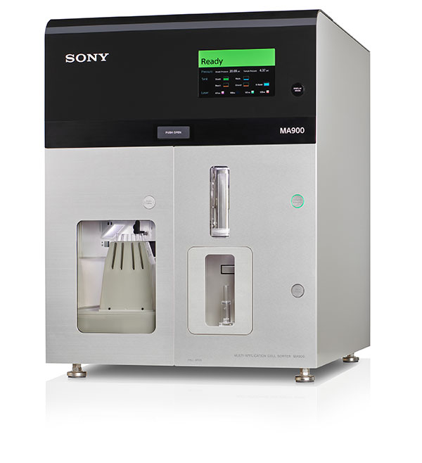
The Sony MA900 Multi-Application Cell Sorter Sharing success is key to unlocking the future of cancer immunotherapy
Cancer treatment is a specialized and evolving field, but the commonality across various histologies and treatments is that many patients relapse and can succumb to the malignancies. Sade-Feldman believes that a global effort in data sharing and transparency is the way of the future in cancer immunotherapy. Sharing protocols and results, across platforms such as the National Institutes of Health’s Gene Expression Omnibus (GEO), would allow specialized techniques and analysis to be tested, replicated, and validated. “[Open access] means that the scientific community can use the latest methods available; we have platforms like the Sony MA900 that make that possible. If we succeed, we want to share our success with others. Eventually, this will lead to an increase in high-quality, robust datasets, which will guide us in making the correct decisions,” concludes Sade-Feldman.
Class 1 Laser Product.
For Research Use Only. Not for use in diagnostic or therapeutic procedures.
References
- Sade-Feldman M, Yizhak K, Bjorgaard SL, et al. Defining T cell states associated with response to checkpoint immunotherapy in melanoma [published correction appears in Cell. 2019;176(1-2):404]. Cell. 2018;175:998-1013.e20. PubMed
- Helmink BA, Reddy SM, Gao J, et al. B cells and tertiary lymphoid structures promote immunotherapy response. Nature. 2020;577:549-555. PubMed