MA900 Multi-Application Cell Sorter

High-purity 4-way sorting of a 12-color immunophenotyping panel

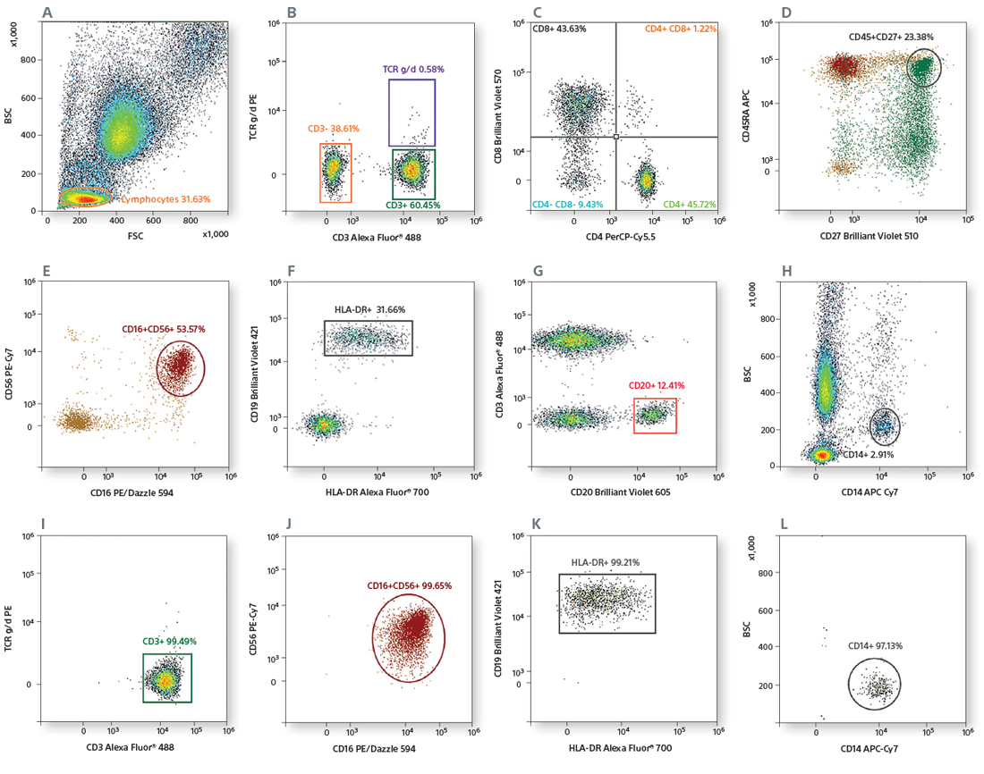
Whole blood was lysed with RBC Lysis Buffer and stained with Alexa Fluor® 488 CD3, PerCP-Cy™5.5 CD4, BD Horizon Brilliant™ Violet 570 (BV570) CD8, APC-Cy™7 CD14, PE/Dazzle™ 594 CD16, BV421 CD19, BV605 CD20, BV510 CD27, APC CD45RA, PE-Cy7 CD56, Alexa Fluor® 700 HLA-DR, and PE TCR gamma/delta antibodies. Cells were incubated for 20 minutes on ice, washed 2X with staining buffer, and analyzed on the MA900 equipped with 488-nm, 405-nm, and 638-nm lasers.

Scatter was used for gating lymphocytes (A). The CD3+ population (B) was used to gate CD4+ and CD8+ cells (C). CD4+ T-cell subsets were identified based on CD45RA and CD27 expression (D). CD16+CD56+ NK cells were gated from CD3- cells (E). CD19+CD20+ B cells were gated from CD3- cells (F), and the HLA-DR expression of B cells was analyzed (G). CD14+ monocytes were identified based on scatter (H). CD3+ T cells, CD19+CD20+ B cells, CD16+CD56+ NK cells, and CD14+ monocytes were sorted by 4-way sorting. Post-sort analysis of each sorted population is shown (I–L).

High-purity 4-way sorting of a 12 color immunophenotyping panel

High-purity sorting of a 12-color immunophenotyping panel using 5 violet fluorochromes

Whole blood was lysed with RBC Lysis Buffer and stained with the following antibodies: Alexa Fluor® CD3, PE CD19, PerCP-Cy5.5 CD8, PE/Dazzle 594 CD16, BD Horizon Brilliant Violet 421 (BV421) CCR7, BV510 CD27, BV605 CD20, BV650 CD45RA, BV711 HLA-DR, and BV750 CD4. Cells were incubated for 20 minutes on ice, washed 2X with staining buffer, and analyzed on the MA900. Lymphocytes were gated using the scatter gate (A).

The CD3+ population (B) was used for gating CD4+ and CD8+ cells (C). CCR7+CD45RA+ lymphocytes are shown in (D). CD19+CD20+ B cells were gated from lymphocytes (E), and the HLA-DR expression of B cells was analyzed (F). CD16+ NK cells (G) were gated from lymphocytes. CD27 expression of CD45RA+ subsets of lymphocytes was analyzed (H).
Using the 4-way sort mode on the MA900 cell sorter, CD4+ and CD8+ T cells, CD19+ B cells, and CD16+ NK cells were sorted. Post-sort analysis of each sorted population is shown (G–J).

Sorting of a 12 color TBNK panel using a 4 laser configuration

High-purity sorting of regulatory T cells

Mouse spleen cells were stained with following antibodies: CD4 PerCP-Cy5.5, Alexa Fluor® 700 CD25, and PE CD127. Cells were incubated for 20 minutes on ice, washed 2X with staining buffer, and analyzed on the MA900. Live spleen cells were gated using the scatter gate (A). The CD4+ population (B) was used to gate CD4+CD25int/highCD127low regulatory T cells (C). These cells were sorted using the MA900. Post-sort analysis of the sorted regulatory T cells is shown (D).

High-purity sorting of regulatory T cells

Cell cycle analysis of Jurkat cells

Jurkat cells were stained using Propidium Iodide (1 µg/mL), and cell cycle analysis was performed using the MA900.

sorting and viability analysis